


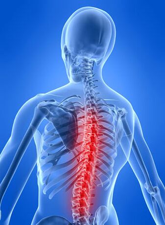
The thoracic form of osteochondrosis is characterized by degenerative damage to the intervertebral cartilage and secondary changes in the thoracic vertebrae. Diagnosis of the disease is sometimes quite problematic, as it is often "masked" as other pathologies: myocardial infarction, angina pectoris, pathologies of the gastrointestinal tract.
Features of thoracic osteochondrosis
This type of disease is quite rare compared to cervical and lumbar.
The reason lies in the peculiarities of the anatomical structure of the thoracic region:
- it is the longest (consisting of 12 vertebrae);
- in this area there is a slight natural bend - physiological kyphosis, which relieves part of the load as a result of upright walking;
- the thoracic region articulates with the ribs and sternum, which perform the functions of a physiological framework and assume the main load;
- in cross section, the spinal canal in the thoracic region has the smallest dimensions;
- The thoracic vertebrae are thinner and smaller in size, but have long spinous processes.
As a result of these factors, the thoracic part is not very mobile, so osteochondrosis in this part of the spine is rare, but its symptoms are quite pronounced: they are quite strong and unpleasant pains associated with pinched spinal nerves, which irritate the shoulder girdle and upper limb organs locatedin the abdominal cavity and chest. For the same reasons, manifestations of the thoracic form of osteochondrosis are often atypical, which significantly complicates the diagnosis of pathology and subsequent treatment.
The narrowness of the spinal canal, the presence of physiological kyphosis and the relatively small size of the vertebrae create the most favorable conditions for the formation of intervertebral disc herniation. Since a significant part of the load falls mainly on the front and lateral parts of the vertebral bodies and discs, the disc is displaced backwards and the formation of a herniated disc, or Schmorl's hernia.
The front part of the vertebrae is exposed to greater stress than the back. For this reason, very often the growth of osteophytes and prolapse of intervertebral discs occurs outside the spine and does not affect the spinal cord.
Stages of thoracic osteochondrosis
Manifestations of thoracic osteochondrosis are determined by changes that occur in the discs and vertebrae, depending on which four main stages of the disease are distinguished:
- Phase I is characterized by dehydration of the intervertebral discs, as a result of which they lose elasticity and firmness, but still retain the ability to withstand normal loads. The process of flattening the disk begins, its height is reduced, and protrusions are formed. Pain at this stage is mild.
- In phase II, cracks form in the fibrous ring and instability of the entire segment is recorded. Painful sensations become more intense and intensify when bending and some other movements.
- A characteristic sign of stage III is rupture of the fibrous ring and the beginning of the formation of a herniated intervertebral disc.
- During the transition to stage IV, due to the lack of resistance from the disc, the vertebrae begin to move closer together, causing spondyloarthrosis (disorders of the intervertebral joints) and spondylolisthesis (twisting or displacement of the vertebrae). The mobilization of compensatory forces to reduce the load leads to growth of the vortex, an increase in its area and flattening. The affected part of the fibrous ring begins to be replaced by bone tissue, which significantly limits the department's motor abilities.
Degrees of thoracic osteochondrosis
Today, many specialists use another classification principle, according to which the course of osteochondrosis of the thoracic spine is distinguished not by stages, but by degrees with their characteristic features.
How does the first degree disease manifest itself? As a rule, it is diagnosed when an intervertebral disc ruptures, caused by overexertion or sudden movement. In this case, a sharp pain in the spine suddenly occurs. Patients compare it to the passage of electric current through the spine. This condition is accompanied by reflex tension of all muscles.
The second degree of thoracic osteochondrosis is spoken about in cases where instability of the spine occurs and symptoms of protrusion of the intervertebral discs develop. This condition is very rare, occurs with periods of exacerbation and subsequent remission, and is discovered only with a thorough diagnostic examination.
What symptoms occur with third degree disease? The pain becomes constant, radiates along the damaged nerve and is accompanied by partial loss of sensation in the upper or lower extremities, changes in gait and intense headache. At this stage, breathing difficulties and disturbance of the normal heart rhythm are often observed.
We can talk about moving to the fourth degree when the manifestations of the disease subside, while the symptoms of spinal instability continue (slipping, twisting of the vertebrae, fixation in relation to each other). Osteophytes begin to grow and gradually squeeze the spinal cord, compressing the spinal cord.
Typical symptoms and signs
Osteochondrosis in the thoracic region has quite characteristic signs, on the basis of which this disease can most likely be diagnosed:
- Intercostal neuralgia - often pain is localized in one area, after which it quickly spreads to the entire chest, which forces patients to be in a certain position and significantly complicates breathing.
- When turning, neck movements, bending, lifting arms, breathing actions (inhalation-exhalation), the pain becomes much more intense.
- The muscles in the middle and upper back go through severe spasms. It is also possible to contract the muscle fibers of the abdominal muscles, lower back and shoulder girdle, which is reflexive in nature (develops as a reaction to a sharp pain syndrome).
- Intercostal neuralgia is often preceded by pain, stiffness and a feeling of discomfort that occurs in the chest and back when moving. The pain can be quite intense and can last for several weeks without spreading further, after which it gradually begins to subside.
- All symptoms become more pronounced at night. In the morning, they soften significantly or subside, intensify with hypothermia, movements (especially vibrating and sudden), and may manifest in the form of a certain stiffness.
Atypical symptoms and signs
Often, the symptoms of osteochondrosis localized in the chest area resemble other diseases.
- Imitation of pain characteristic of cardiac pathologies (heart attack, angina). Such pain can be quite long-lasting (unlike cardialgia), while traditional drugs used to dilate coronary vessels do not eliminate pain. The cardiogram shows no changes either.
- In the acute stage of thoracic osteochondrosis, long-lasting (up to several weeks) soreness in the sternum, reminiscent of diseases of the mammary glands, often occurs. They can be ruled out by examination by a mammologist.
- Pain in the abdomen (iliac region) resembles colitis or gastritis. When localized in the right hypochondrium, cholecystitis, pancreatitis or hepatitis are often misdiagnosed. Such symptoms are often accompanied by disruption of the digestive system due to damage to their innervation. In such cases, it is necessary to identify thoracic osteochondrosis as the primary disease that provokes such manifestations.
- If the lower thoracic region is damaged, the pain is concentrated in the abdominal cavity and simulates intestinal pathologies, but there is no correlation with the quality of food taken and diet. The severity of pain increases mainly due to physical activity.
- Disorders in the reproductive or urinary system also develop as a result of distortion of the innervation of organs.
- Damage to the upper segment of the thoracic region leads to the appearance of symptoms such as pain in the esophagus and pharynx and the sensation of a foreign body in the pharyngeal cavity or in the retrosternal region.
Atypical symptoms are characterized by manifestation in the late afternoon, absence in the morning and occurrence when provoking factors occur.
Dorsago and dorsalgia
Signs of thoracic osteochondrosis include two vertebral syndromes:
- dorsago;
- dorsalgia.
Dorsago is a sudden sharp pain that occurs in the thoracic region, mainly when standing up after a long period of sitting in a bent position. The intensity of the pain can be so high that the person has difficulty breathing. In this case, there is significant muscle tension and limited range of motion in two sections: cervicothoracic and thoracolumbar.
Dorsalgia is characterized by a gradual, imperceptible development. The severity of the pain is small - sometimes it is better to talk about a feeling of discomfort than a pain syndrome. Highlights:
- duration can be up to 14-20 days;
- intensification of the syndrome is observed when bending to the sides, forward or taking a deep breath;
- with upper dorsalgia, movements in the cervicothoracic region are limited, with lower dorsalgia, movements in the lumbar-thoracic region are limited;
- the pain intensifies at night and may completely disappear when you walk;
- increased pain is caused by deep breathing and prolonged stay in one position.
Diagnostics
To confirm the diagnosis, the following is performed:
- Radiography. With its help you can discover:
- changes in the anatomy of the damaged segment;
- thickening of the disc;
- vertebral deformation and displacement;
- difference in the height of intervertebral discs.
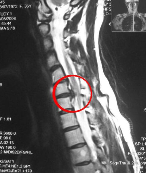
- Computed tomography (CT) and magnetic resonance imaging (MRI) are more accurate methods because they provide a layer-by-layer picture of the affected area.
- Electromyography is performed to differentiate neurological symptoms that develop as a result of compression of the nerve roots in the thoracic type of osteochondrosis. An examination is prescribed if the following signs are present:
- decreased coordination of movements;
- headache;
- dizziness;
- pressure fluctuations.
- Laboratory tests - performed to determine the level of calcium in the blood and ESR (erythrocyte sedimentation rate).